Virtual Museum of Canada

WATERS OF LIFE

LIFE UNDERWATER

BENTHOS - BIODIVERSITY

Top of page Top of page Top of page Top of page Top of page Top of page Top of page Top of page Top of page Top of page Top of page Top of page Top of page Top of page Top of page Top of page Top of page Top of page Top of page Top of page

Get Flash PlayerFLASH PLAYER IS REQUIRED TO SEE THE CONTENT OF THIS PAGE

This page contains videos that require Javascript and Adobe Flash Player to be activated. If you wish not to activate these functions, we offer pictures from the original videos.

Video of zebra mussels underwater

VIDEO - 0 min 13s
Zebra mussels and quagga mussels are invasive exotic species that spread very rapidly.
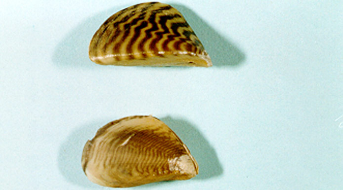
A zebra mussel on top and a quagga mussel in the bottom of the photo

Zebra mussel (up)
quagga mussel (down)

Their presence in the St. Lawrence River was first reported in the 1990s. These mussels attach themselves to hard surfaces, and can cover them completely. By filtering up to one litre of water per day to feed on plankton, they reduce the amount of nutrients available for other organisms. This competition for resources most strongly affects other species of mussel. These mussels disrupt the ecological balance and cause damage by blocking industrial, domestic and municipal water treatment plant intake structures.

Gastropods on a water-lily leaf

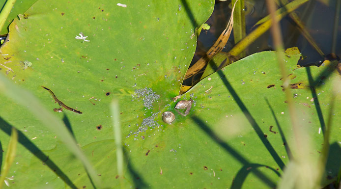
Gastropod molluscs have only one shell. Different kinds of gastropods are present in Lake Saint-Pierre.

Top of page

This page contains videos that require Javascript and Adobe Flash Player to be activated. If you wish not to activate these functions, we offer pictures from the original videos.

Video filmed under a microscope of a Physidae

VIDEO - 0 min 15 s
The Physidae have a spiral shell with a left aperture.

This page contains videos that require Javascript and Adobe Flash Player to be activated. If you wish not to activate these functions, we offer pictures from the original videos.

Video filmed under a microscope of a Planorbidae

VIDEO - 0 min 15 s
The Planorbidae have a shell that is coiled flat rather than being conical.

This page contains videos that require Javascript and Adobe Flash Player to be activated. If you wish not to activate these functions, we offer pictures from the original videos.

Video filmed under a microscope of an Hydrobiidae

VIDEO - 0 min 12 s
The Hydrobiidae have a shell with a right aperture.

Top of page

This page contains videos that require Javascript and Adobe Flash Player to be activated. If you wish not to activate these functions, we offer pictures from the original videos.

Video of an Lymnaeidae in an aquarium

VIDEO - 0 min 12 s
The same is true of the Lymnaeidae.

This page contains videos that require Javascript and Adobe Flash Player to be activated. If you wish not to activate these functions, we offer pictures from the original videos.

Video filmed under a microscope of a dragonfly larva and a damselfly larva

VIDEO - 0 min 10 s
Many types of insect larvae also live in the water.
Odonates are divided into two groups: Zygoptera (damselflies) and Anisoptera (dragonflies). These formidable predators have very large eyes.

This page contains videos that require Javascript and Adobe Flash Player to be activated. If you wish not to activate these functions, we offer pictures from the original videos.

Video filmed under a microscope of an odonate anisoptera

VIDEO - 0 min 34 s
Odonate larva, Anisoptera

Top of page

This page contains videos that require Javascript and Adobe Flash Player to be activated. If you wish not to activate these functions, we offer pictures from the original videos.

Video filmed under a microscope of an odonate zygoptera

VIDEO - 0 min 23 s
Odonate larva, Zygoptera

This page contains videos that require Javascript and Adobe Flash Player to be activated. If you wish not to activate these functions, we offer pictures from the original videos.

Video filmed under a microscope of a caddisfly in a case made of vegetable debris

VIDEO - 0 min 18 s
Trichopterans, or caddisflies, are true artisans that build a case for themselves out of vegetable or mineral debris. The case is held together by silk secreted from their salivary glands. Caddisfly cases come in a variety of shapes.

This page contains videos that require Javascript and Adobe Flash Player to be activated. If you wish not to activate these functions, we offer pictures from the original videos.

Video filmed under a microscope of a caddisfly without a real case

VIDEO - 0 min 15 s
Other caddisflies have no real case but produce structures of silk to filter or trap their prey.

Top of page

This page contains videos that require Javascript and Adobe Flash Player to be activated. If you wish not to activate these functions, we offer pictures from the original videos.

Video filmed under a microscope of a caddisfly capturing its food

VIDEO - 0 min 10 s
These silk structures are less developed than the cases but they are used to capture food.

This page contains videos that require Javascript and Adobe Flash Player to be activated. If you wish not to activate these functions, we offer pictures from the original videos.

Video filmed under a microscope of a caddisfly in a case made of plant materials

VIDEO - 0 min 08 s
Others use plant materials to build themselves highly elaborate cases.

This page contains videos that require Javascript and Adobe Flash Player to be activated. If you wish not to activate these functions, we offer pictures from the original videos.

Video filmed under a microscope of an odonate zygoptera

VIDEO - 0 min 08 s
We observe in this video an odonate larva, Zygoptera.

Top of page

This page contains videos that require Javascript and Adobe Flash Player to be activated. If you wish not to activate these functions, we offer pictures from the original videos.

Video filmed under a microscope of an ephemeropteran

VIDEO - 0 min 08 s
Ephemeropterans, or mayflies, generally have three tails, caudal filaments, on the tip of their abdomen.

This page contains videos that require Javascript and Adobe Flash Player to be activated. If you wish not to activate these functions, we offer pictures from the original videos.

Video filmed under a microscope of an ephemeropteran and a Dytiscidae

VIDEO - 0 min 07 s
This video shows a mayfly larva and a Dytiscidae larva.

This page contains videos that require Javascript and Adobe Flash Player to be activated. If you wish not to activate these functions, we offer pictures from the original videos.

Video filmed under a microscope of a beetle adult

VIDEO - 0 min 12 s
Some families of beetles (Dytiscidae, Haliplidae, Gyrinidae), live in the water from the larval stage to the adult stage. In the adults, the front wings, or elytra, are hard and help make the organism more waterproof.

Top of page

This page contains videos that require Javascript and Adobe Flash Player to be activated. If you wish not to activate these functions, we offer pictures from the original videos.

Video filmed under a microscope of a beetle larva

VIDEO - 0 min 12 s
This beetle larva has various filaments on its body.

This page contains videos that require Javascript and Adobe Flash Player to be activated. If you wish not to activate these functions, we offer pictures from the original videos.

Video filmed under a microscope of a Dytiscidae larva

VIDEO - 0 min 16 s
Dytiscid larvae come in various shapes.

This page contains videos that require Javascript and Adobe Flash Player to be activated. If you wish not to activate these functions, we offer pictures from the original videos.

Video filmed under a microscope of a beetle

VIDEO - 0 min 08 s
This beetle is in the family Haliplidae.

Top of page

This page contains videos that require Javascript and Adobe Flash Player to be activated. If you wish not to activate these functions, we offer pictures from the original videos.

Video filmed under a microscope of an hemipteran

VIDEO - 0 min 07 s
The wings of hemipterans are horny at the base and membranous at the tips.

This page contains videos that require Javascript and Adobe Flash Player to be activated. If you wish not to activate these functions, we offer pictures from the original videos.

Video filmed under a microscope of an hemipteran of the Pleidae family

VIDEO - 0 min 10 s
This adult hemipteran is in the family Pleidae.

This page contains videos that require Javascript and Adobe Flash Player to be activated. If you wish not to activate these functions, we offer pictures from the original videos.

Video filmed under a microscope of an hemipteran of the Corixidae family

VIDEO - 0 min 17 s
This other hemipteran is in the family Corixidae.

Top of page

This page contains videos that require Javascript and Adobe Flash Player to be activated. If you wish not to activate these functions, we offer pictures from the original videos.

Video filmed under a microscope of an hemipteran of the Nepidae family

VIDEO - 0 min 07 s
This large hemipteran in the family Nepidae, commonly known as a water scorpion, uses the hooks on its front feet to hold its prey.

This page contains videos that require Javascript and Adobe Flash Player to be activated. If you wish not to activate these functions, we offer pictures from the original videos.

Video filmed under a microscope of a brown hydracarina

VIDEO - 0 min 18 s
Water mites (Hydracarina) are generally under 4 mm long. They can move rapidly on their four pairs of articulated legs, which reveal their kinship with spiders.

This page contains videos that require Javascript and Adobe Flash Player to be activated. If you wish not to activate these functions, we offer pictures from the original videos.

Video filmed under a microscope of a red hydracarina

VIDEO - 0 min 15 s
Several hypotheses have been advanced to explain the red pigmentation of this water mite. Some believe this bright colour, which is easily seen by fish, marks the water mite as being inedible because of its bad taste.
Other scientists believe that this coloration is an adaptation to help capture solar energy in cold water.

Top of page

This page contains videos that require Javascript and Adobe Flash Player to be activated. If you wish not to activate these functions, we offer pictures from the original videos.

Video filmed under a microscope of a white planarian

VIDEO - 0 min 29 s
Planarians are very common in fresh water. They can be recognized by their flat body and their head, which appears to have two ears and two eyes.

This page contains videos that require Javascript and Adobe Flash Player to be activated. If you wish not to activate these functions, we offer pictures from the original videos.

Video filmed under a microscope of a brown planarian

VIDEO - 0 min 13 s
These are in fact primitive eyes, known as ocelli, and allow the worm to move away from light sources.

This page contains videos that require Javascript and Adobe Flash Player to be activated. If you wish not to activate these functions, we offer pictures from the original videos.

Video filmed under a microscope of an oligochaete

VIDEO - 0 min 12 s
Oligochaetes, a type of worm, have bristles growing from their segmented bodies. They are related to earthworms. They live on the bottom of lakes and rivers and sometimes swim within the water column. They sift through the sediments, feeding on debris, decomposing matter and algae.

Top of page

This page contains videos that require Javascript and Adobe Flash Player to be activated. If you wish not to activate these functions, we offer pictures from the original videos.

Video filmed under a microscope of a nematod

VIDEO - 0 min 25 s
Nematodes are small round worms less than 10 centimetres long. Their smooth bodies are pointed at both ends.

This page contains videos that require Javascript and Adobe Flash Player to be activated. If you wish not to activate these functions, we offer pictures from the original videos.

Video filmed under a microscope of an hydra

VIDEO - 0 min 17 s
The hydra is a predator that is attached to the substrate. This cnidarian captures zooplanktonic organisms by injecting a toxin that paralyzes them. When a hydra is threatened, it retracts its tentacles.

This page contains videos that require Javascript and Adobe Flash Player to be activated. If you wish not to activate these functions, we offer pictures from the original videos.

Video filmed under a microscope of an ostracod

VIDEO - 0 min 20 s
Ostracods are shaped like mussels, although in fact they are small crustaceans under 4 mm long. Their head and body are completely enclosed in two valves that open in the front. Only the animal’s appendages emerge from the shell, allowing it to move around.

Top of page

This page contains videos that require Javascript and Adobe Flash Player to be activated. If you wish not to activate these functions, we offer pictures from the original videos.

Video filmed under a microscope of an amphipod

VIDEO - 0 min 10 s
Amphipods are small crustaceans that are common in Lake Saint-Pierre. They can be recognized by their laterally flattened bodies and resemble shrimp.

This page contains videos that require Javascript and Adobe Flash Player to be activated. If you wish not to activate these functions, we offer pictures from the original videos.

Video filmed under a microscope of an isopod

VIDEO - 0 min 14 s
Isopods, another type of crustacean, are flattened dorso-ventrally. They move on or within the substrate. Aquatic isopods, like sow bugs on land, feed on vegetables and animals, sometimes injured or dead.

Face view of the Chinese mitten crab

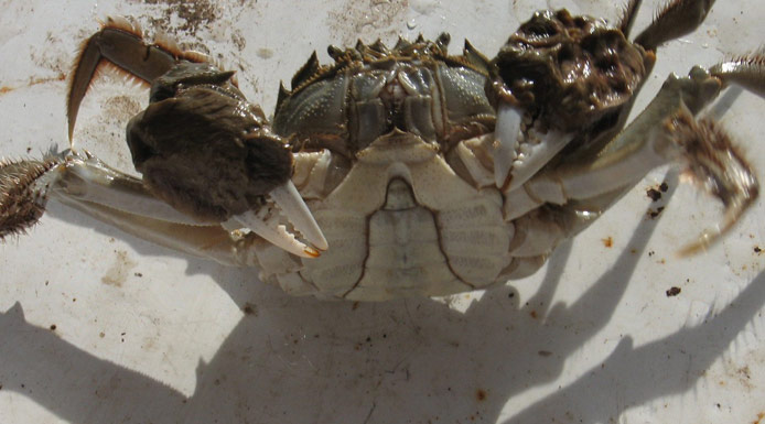
The Chinese mitten crab is an invasive exotic species that was first observed in the St. Lawrence in 2004, near Quebec City.

Top of page

Chinese mitten crab seen from below

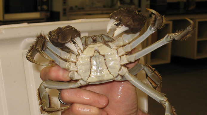
In 2005 and 2006, specimens were found in Lake Saint-Pierre.

Chinese mitten crab showing his claws

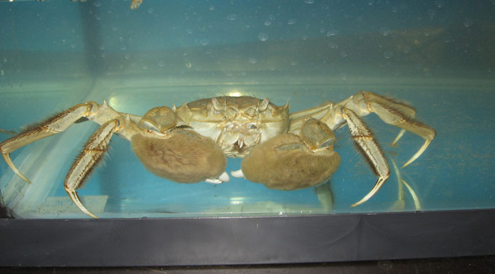
With its capacity to rapidly colonize new habitats, this crab could threaten local biodiversity and ecosystem functioning.

Top of page

LIFE UNDERWATER

BENTHOS